NEET Biology Notes Biological Classification
Five Kingdom Classification
RH Whittaker (1969) propositi a five kingdom classification. The kingdom names were Monera, Protista, Fungi, Plantae and Animalia.
The main criteria used by him for classification were cell structure, body organisation, mode of nutrition, reproduction and phylogenetic relationship.

Kingdom-Monera
Kingdom-Monera includes all prokaryotic organisms, i.e. bacteria, mycoplasma, actinomycetes, spirochaetes, rickettsiae, chlamydiae and cyanobacteria.
Features of Kingdom-Monera
Members of this kingdom are known as monerans, which lack true nucleus, nuclear membrane, nucleolus, chromatin, histone proteins and all membrane bound organelles.
Genetic material is called nucleoid or genophore or incipient nucleus or prochromosome, which is composed of naked DNA, RNA and proteins.
Plasmids are small extrachromosomal rings of DNA discovered by Hayes and Lederberg (1952). They can replicate independently without nucleoid. If associated with nucleoid they are called episomes. Ribosomal RNA are of 70 S type. Cell wall is made up of peptidoglycan (except archaebacteria and mycoplasma). Reproduction is usually asexual by means of binary fission.
Respiratory enzymes are found associated with the plasma membrane. Cell division is amitotic type and lacks spindle formation. Two major bacterial lineages are recognised, i.e. the primitive archaebacteria and more advanced eubacteria.
The split leading to the arcftaea and the eukaryotes occurred after the bacteria had gone their own way.

However, the acquisition of mitochondria by eukaryotes (probably from an ancestor of today’s rickettsias) and chloroplasts (from cyanobacteria) occurred after their line had diverged from the archaea.
Bacteria
Bacteria are microscopic, relatively simple, unicellular organism, whose genetic material is not enclosed in nuclear membrane. These are cosmopolitan in distribution. Bacteria are of two types on the basis of structure and evolution namely archaebacteria and eubacteria.
Archaebacteria
It is a primitive group of bacteria. These are oldestof the living fossils. Archaebacteria inhabit mostextreme environments on earth that’s why, they arecalled extremophiles.
Features of Archaebacteria
- Their cell walls lack peptidoglycan which is an important component of the cell wall of eubacteria.
- The lipids contain phytanyl side chains (branched lipid). Branch chained lipids decrease membrane fluidity and increase tolerance to extremes of heat as well as low pH.
- The presence of distinctive ribosomal RNA sequences.
- The presence of introns (i.e. non-coding genes) like that of eukaryotes.
Groups of Archaebacteria
The three main groups of archaebacteria are :
- Methanogens These are obligate anaerobic (i.e. free oxygen will kill them), Gram negative bacteria, which utilise C02 during cellular respiration to produce methane (CH4) as a waste product.
These are found in the musk of swamps and marshes, the rumen of cattle (where they live on the hydrogen and C02 produced by other microbes living along with them), our colon (large intestine), sewage sludges and the gut of termites. These are chemoautotrophs, which use hydrogen as a source of electrons for reducing carbon dioxide to food and giving off methane (marsh gas andCH4) as a byproduct.
4H2 + C02—- > CH4 + 2H20
e.g. Methanobacterium, Methanobacillus, Methanosarcina, Methanococcus
- Halophiles These are found in extremely saline environments such as great salt lake in the US and Dead sea. They maintain osmotic balance with their surroundings by building up the solute concentration within their cells.
- Halophiles (anaerobic bacteria) trap solar energy by purple pigmented membrane to produce ATP, e.g. Haloarcula, Halobacterium, etc.
- Thermoacidophiles These are found in hot and acidic places such as acidic sulphur springs (i.e. in Yellowstone National Park) and deep-sea-vents (black smockers).
- Some forms of thermoacidophiles (aerobic bacteria) reduce sulphur to H2S under anaerobic conditions, e.g. Sulfolobus, Thermoproteus, etc
- Phylogenitically, it is considered that archaebacteria are more close to eukaryotes than bacteria because archaebacteria have mix traits of bacteria as well as traits found in eukaryotes

Eubacteria
- They are typical prokaryotic (i.e. no membrane encloses nucleus) cells with a single chromosome (i.e. a closed circle of double stranded DNA, without histone protein) and without mitochondria, chloroplast and any other membrane bound cell organelles. These are cosmopolitan in distribution, which are found in water, soil, air and in plant and animal body.
- The size of bacteria ranges from 0.1-1.5 pm in diameter and 2-10 pm in length. Dialister pneumosintes (0.15-0.3 pm long) is the smallest bacterium, while Beggiatoa mirabilis (16-45 pm diameter and up to several centimeter long) is the largest bacterium.
Shape of Eubacteria
- On the basis of shape, bacteria can be either coccus (i.e. spherical or nearly spherical, small and non-flagellated), bacillus (i.e. rod-shaped, e.g. Escherichia coli and Lactobacillus), vibrio (i.e. comma-shaped, e.g. Vibrio cholerae), spiral, i.e. coiled form of bacteria exhibiting twist with one or more turns, e.g. Spirillum minus.
- Coccus bacteria can be either micrococcus (occurs singly, e.g. Micrococcus agilis, Micrococcus roseus) Diplococcus (found in pairs.
e.g. Gonococcus, Meningococcus, Diplococcus pneumoniae).
Streptococcus (i.e. cells remain attached to form a chain.e.g. Streptococcus lactis), staphylococcus irregular bunches of cells or grape-like clusters,
e.g. Staphylococcus aureus and sarcinae, cubical packets of 8; 27 or more cells
e.g. Sarcina.Bacilli bacteria arranged in pairs are called diplobacilli e.g. bacilli arranged in chain are called streptobacilli, e.g. Streptobacillus, Azotobacter, Bacillus anthracis, bacilli arranged side-by-side like matchstics are called palisade like, e.g. Coiynebacterium diphtheriae, when the cells of a chain have much larger area of contact with each other, it is called trichomes
e.g. Biggiatoa. Caulobacter is stalked bacterium, Rhodomicrobium is budding bacterium, while Bhizobium, Corynebacterium, Azotobacter and Mycobacterium are called pleomorphic bacteria.
Structure of Bacterial Cell
Structural components of bacteria are as follows :
Bacterial Flagella
In bacteria, flagella may or may not be present. If present they are made up of a single filament of the protein flagellin, instead of 9 + 2 tubulin containing microtubules found in eukaryotes. It consists of basal body, hook and filament. Basal body is the most complex portion of flagellum having 4 rings (L, P, S and M) in Gram negative and 2 rings (S and M) in Gram positive bacteria. Hook consists of a single type of protein and functions to connect the filament to motor portion, while filament is the longest portion of flagellum, which is 20 nm wide and 1-70 nm long and consists of 8 vertical rows of flagellin.
Flagellar Arrangement
Bacterial cells have following types of flagellar arrangement:
- Atrichous Flagella absent, e.g. Lactobacillus, Pasteurella.
- Monotrichous Single polar flagellum, e.g. Vibrio cholerae and Thiobacillus.
- Amphitrichous Single flagellum at each end of the cell, e.g. Nitrosomonas.
- Cephalotrichous Bacteria with many flagella attached at one end, e.g. Pseudomonas fluorescence.
- Lophotrichous Bacteria with a group of flagella attached at each end, e.g. Spirillum volutans.
- Peritrichous Bacteria with flagella all over the body, e.g. E. coli and Clostridium tetani.
Pilin or Fimbriae
These are extremely minute hair-like structures found mostly in male bacterial cells. Fimbriae are shorter and thinner (3-10 nm diameter) than flagellum and used for attachment. They are composed of protein sub-units called pilin. A special type of pili called sex pili present on male bacterium makes conjugation tube during conjugation.
Cell Wall
- It is the outer tough covering around the cell, which provides specific shape and protection to the bacterium. It prevents the cell from swelling and bursting due to the osmotic changes.
- It is composed of a polysaccharide called murein or peptidoglycan. It consists of polysaccharide cross linked with short amino acid chains.
- The cell wall contains some special amino acids such as L-alanine, D-alanine, D-glutamic acid and either lysine or diaminopimelic acid. Antibiotics such as penicillin, cephalosporin, etc., prevent the cross linking and inhibit the bacterial cell wall formation.
- Lysozyme attacks on the bond between glucosamine and muramic acid and hydrolyses the cell wall. The cell wall of Gram positive bacteria is much thicker and contains less lipids compared to that of Gram negative bacteria. The cell wall can be dissolved by the enzyme lysozyme, an antibacterial enzyme found in tears, saliva, egg white and some other body fluids.
- Lysozyme digests the’polysaccharide back bone of murein.
- Lipopolysaccharides (LPS) are found in outer membrane of cell wall in Gram negative bacteria. It acts as surface antigen in cell wall.
Cytoplasm
It is a semi-fluid ground substance enclosed by the plasma membrane. Plasma membrane is a phospholipid bilayer but there is no cholesterol or other steroids. Cytoplasm appears granular due to the presence of large number (as many as 20,000) of 70 S type ribosomes, which may occur singly or in clusters called polyribosome.
Apart from the nucleoid, certain bacterial cells contain extrachromosomal genetic material called plasmids. The plasmid DNA replicates independently. It contains genes like fertility factor (F-factor), resistance factor (R-factor), nitrogen fixing genes (Nif-genes). Some plasmids may temporarily become associated with nucleoid DNA and are known as episomes.
Plasmid term was coined by Lederberg and Hayes. Each plasmid is circular, supercoiled, double stranded and naked DNA.
Staining of Bacteria
The Gram staining based on cell wall structure is named by Danish bacteriologist Christian Gram, who developed it in 1884. Bacteria that are not decolourised by the alcohol/acetone wash are Gram positive. The bacterial cells are first stained with a purple dye called crystal violet. Then the preparation is treated with alcohol or acetone.

To see them now requires the use of a counterstain of a different colour (e.g. the pink of safranin).
Gram Positive Bacteria
- Not as diverse as the Gram negative bacteria, the Gram positive still make up an impressively varied group.
- This division includes the Gram positive rods, Gram positive cocci and the Actinomycetes, which exhibit superficial similarity and function (but not evolutionary).
Gram Negative Bacteria
- About 75% of known eubacteria are Gram negative.
- Gram negative bacteria form an extremely diverse group. The fact all Gram negative does not necessarily imply that they comprise a monophyletic taxon.

Nutrition in Bacteria
Bacteria exhibit different modes of nutrition. On this basis, broadly two types of bacteria can be recognised; autotrophic bacteria and heterotrophic bacteria.
(i) Autotrophic Bacteria
These are able to synthesise their own organic food from inorganic substances. They use carbon dioxide for obtaining carbon and utilise hydrogen sulphide (H2S) or ammonia (NH3) or hydrogen (H2) as the source of hydrogen to reduce carbon. These bacteria can be distinguished further into two types, i.e. photoautotrophic bacteria and chemosynthetic bacteria.
(a) Photoautotrophic Bacteria
These possess photosynthetic pigments in membrane bound lamellae (thylakoids) and utilise solar energy. The bacterial photosynthesis is different from that of green plants since, here water is not used as a hydrogen donor. Hence, oxygen is not released as a byproduct. For this reason, the process is described as anoxygenic photosynthesis.
Light energy
CO2 +H2S————- > Sugar + Sulphur + Water
- Chemosynthetic Bacteria
The bacteria, which manufacture organic compounds from inorganic raw materials utilising energy, liberated from the oxidation of inorganic substances.
The following are the common types of chemoautotrophic bacteria :
- Nitrifying bacteria Which derive energy by oxidising ammonia into nitrates,
e.g. Nitrosomonas and Nitrobacter.
NH4+202 ——- > N02 +2H20 + energy
- Sulphur bacteria Which derive energy by oxidising hydrogen sulphide to sulphur,
e.g. Thiobacillus and Beggiatoa.
2H2S + 02 —– > 2S + 2H20 + energy
- Iron bacteria Which derive energy by oxidising ferrous ions into ferric form, e.g. Ferrobacillus and
4FeC03 +6H20 + 02 —> 4Fe (OH)3
+4C02 + energy
(ii) Heterotrophic Bacteria
These are unable to manufacture their own organic food and hence, are dependent on external source. These bacteria can be distinguished into saprophytic bacteria, symbiotic bacteria and parasitic bacteria.
- Saprophytic Bacteria
- They obtain their nutritional requirements from dead organic matter. They breakdown the complex organic matter into simple soluble form by secreting exogenous enzymes. Subsequently, they absorb the simple nutrients and assimilate them, during which they release energy. These bacteria have a significant role in the ecosystem, functioning as decomposers.
- The aerobic breakdown of organic matter is called as decay or decomposition. It is usually complete and not accompanied by the release of foul gases. Anaerobic breakdown of organic matter is called fermentation. It is usually incomplete and is always accompanied by the release of foul gases. Anaerobic breakdown of proteins is called putrifaction.
- The property of decomposition of organic compounds is employed in several industrial processes such as ripening of cheese, in the retting of fibres and in the curing of tobacco.
- Symbiotic Bacteria
They live in a mutually beneficial association with other organisms. Such bacteria derive the essential nutrients from their host organisms and in that process help the host through some of their biological activities.
The most familiar example of symbiotic bacteria are the nitrogen fixing bacteria, found in the root nodules of leguminous plants. Bacteria such as Rhizobium and Pseudomonas reside in the root nodules and reduce atmospheric nitrogen directly to ammonia. This becomes the source of nitrogen for the host plants. The plants in return provide bacteria with nutrients and protection.
Nitrogenase enzyme system helps in fixation of nitrogen. This system is made up of two components, i.e. nitrogenase and nitrogenase reductase. Blue-green algae (cyanobacteria) fix nitrogen in heterocysts under anaerobic conditions. Rhizobium bacteria needs leghaemoglobin fixation of nitrogen. Nif gene is responsible for synthesis of these enzyme.
This gene is found in plasmids of prokaryotes. Because plasmids are not found in eukaryotes, they cannot fix atmospheriq nitrogen.
The bacteria found in the human alimentary canal, Escherichia coli are non-pathogenic. These bacteria check the growth of harmful putrefying bacteria. In addition, these bacteria release vitamin-K an$ B12, which are necessary for blood components. The human host provides shelter and food for these bacteria.
A similar example is that of cellulose digesting bacteria, which occur in the alimentary canal of ruminant mammals such as cows and goats.
- Parasitic Bacteria
This bacteria occur in the body of animals and plants, obtaining their organic food from there. Most of these bacteria are pathogenic, causing serious diseases in the host organisms either by exploiting them or by releasing poisonous secretion called toxins. Toxins are of two types i.e; exotoxins and endotoxins.

Respiration in Bacteria
Based on mode of respiration, bacteria may be
- Obligate aerobic bacteria Which can perform only aerobic respiration, e.g. Bacillus subtilis.
- Obligate anaerobic bacteria Which can perform only anaerobic respiration, e.g. Clostridium botulinum.
- Facultative aerobes Which are anaerobic bacteria, but can live in the presence of oxygen, e.g. Chlorobium.
- Facultative anaerobes Which are aerobic bacteria, but can live in the absence of oxygen also, e.g. Pseudomonas.
Reproduction in Bacteria
These are of the following three types :
- Vegetative Reproduction
It takes place by binary fission, in which the bacterial DNA undergoes replication and under favourable conditions the bacterial cell expands and cytoplasm divides into two parts. - Asexual Reproduction
It takes place by endospore formation.
Bacterial population is counted by 2n[n = number of generation). For example, a bacterial cell after 5 generations of fission will resulted into 25 = 2x2x2x2x2=32 bacterial cells. Some bacteria produce non-motile spores such as conidia (e.g. Streptomyces), oidiospores (e.g .Actinomyces), endospores (e.g. Bacillus) etc. - Sexual Reproduction
It takes place by genetic recombination. There are three methods of genetic recombination in bacteria. - Transformation
In transformation, DNA released from one bacterial cell is taken up by another without any contact between donor and recipient bacterium. It was discovered by Griffith in 1928, when he was working with the bacterium Diplococcus pneumoniae or Pneumococcus pneumoniae, which causes pneumonia. In 1944, Avery, MacLeod and McCarty succeeded in isolating and identifying the transforming factor and found that it was DNA. This was the first direct evidence that DNA is the genetic material. - Conjugation
In conjugation, transfer of DNA involves direct cell to cell contact and a conjugative plas’mid in donor cell. Conjugation was discovered by Lederberg and Tatum (1946) in E. coli.
Conjugation occurs between donor cell (F+) and recipient cell (F_). Donor cell is having sex pili and F-factor, whereas, recipient cell is lacking both. Conjugation may involve either only the replication and transfer of the F-plasmid (fertility factor or F-factor) from donor (F+) to the recipient (F“), thus making the later also a donor or integration of F-plasmid with the chromosome of donor to form episome.
Term ‘episome’ is applied to extra-nuclear genetic material, which may remain integrated or free state, e.g. F-factor, temperate phage, etc. All plasmids are episomes but all episomes are not plasmids.
When F-factor is integrated with main genome or nuclear DNA, the frequency of recombination increases by 1000 times. That is why, the donor cell is called Hfr-donor (High frequency donor) or high frequency male. - Transduction
In transduction, transfer of genetic material between bacteria is mediated by viruses, e.g. bacteriophage. Transduction was first of all reported in Salmonella typhimurium by Zinder and Lederberg (1952).
Types of Transduction
Generalised transduction, i.e. transducing bacteriophage can transfer any gene of the donor bacterium, e.g. T4-bacteriophage.
Restricted (specialised) transduction, i.e. transducing bacteriophage can carry only a specific region of the bacterial DNA to a recipient
e.g. bacteriophage.
Abortive transduction, i.e. DNA fragment from donor bacterium is not integrated in the genome of recipient bacterium and is lost after one or few generations.
Some Popular Bacteria
Some popular bacteria are as follows:
- (i) Rickettsias
These are obligate intracellular parasites. This means that they can only grow and reproduce, while within the living cells of their host including certain arthropods (ticks, mites, lice and fleas) and mammals.
Rickettsia prowazekii causes typhus fever, when it is transmitted to humans by lice. Rocky mountain spotted fever is a rickettsial disease transmitted by ticks.
The mitochondria of eukaryotes probably evolved from endosymbiotic bacteria. Because of the similarities of their genomes, rickettsias may be the closest relatives to the ancestors of mitochondria. - Nitrosomonas
This chemoautotroph oxidises NH3 (produced from proteins by decay bacteria) to nitrites (N02). This provides the energy to drive their anabolic reactions. The nitrites are then converted (by other nitrifying bacteria) into nitrates (NO 3~), which supply the nitrogen needs of plants. - Neisseria meningitis
It causes meningococcal meningitis, an extremely serious infection of the meninges that occasionally occurs in very young children and in military camps. Neisseria gonorrhoeae causes gonorrhoea, one of the most common Sexually Transmitted Diseases (STDs). - Escherichia coli
It is most thoroughly-studied of all creatures (possibly excepting ourselves). Its entire genome has been determined down to the last nucleotide 4,639,221 base pairs of DNA encoding 4,377 genes. It lives in the human colon, usually harmlessly. However, water or undercooked food contaminated with the Ol57:H7 strain has caused severe and occasionally fatal infections. - Salmonella enterica
Two major human pathogens are as follows :
(a) Salmonella enterica var. typhi causes typhoid fever, a serious systemic infection occurring only in humans. This microbe is also known as Salmonella typhi.
(b) Salmonella enterica var. typhimurium confined to the intestine, it is a frequent cause of human gastrointestinal upsets but is also found in many other animals (that are often of the source of she human infection). Also known as Salmonella typhimurium. - Vibrio cholerae
It causes cholera, one of the most devastating of- the intestinal diseases. The bacteria liberate a toxin that causes massive diarrhoea (10-15 L/day) and loss of salts. Unless the water, and salts are replaced quickly, the victim may die (of shock) in a few hours. Like other intestinal diseases, cholera is contracted by the ingestion of food or more often, water that is contaminated with the bacteria. - Yersinia pestis
This bacillus causes bubonic plague. It is usually transmitted to human by the bite of an infected flea. As it spreads into the lymph nodes, it causes them to become greatly swollen, hence the name ‘bubonic’ (bubo-swelling of a lymph node) plague. Once in the lungs, however, the bacteria can spread through the air causing the rapidly lethal (2-3 days) ‘pneumonic’ plague. Untreated, 50-75% of the cases of bubonic plague are fatal and figure for the pneumonic form reaches 100%.
- Purple Sulphur Bacteria .
Like green plants, these are photosynthetic, using the energy of sunlight to reduce carbon dioxide to carbohydrate. Unlike plants however, they do not use water as a source of electrons. Instead, they use hydrogen sulphide to supply the electrons needed to synthesise NADPH and ATP.
2H2S + C02 —> (CH20) -l-H20 + 2S
In the process, they produce elemental sulphur (e.g. Chromatium). Photosynthetic bacteria contain special types of chlorophylls called bacterio chlorophylls incorporated into membranes. With this machinery, they can run photosystem-I but not photosystem-II (which explains their inability to use water as a source of electrons).*
Most photosynthetic bacteria are obligate anaerobes. They cannot tolerate free oxygen. Thus, they are restricted to such habitats as the surface of sediments, at the bottom of shallow ponds and estuaries. However, the absorption spectrum of their bacteriochlorophylls lies mostly in the infra-red region of the spectrum so they can trap energy missed by the green plants above them.
- Bacillus sp.
These organisms differ mainly in the plasmids they contain B. anthrr causes anthrax. Currently, the biological agent b„mg used by terrorists. Its two plasmids contain the genes needed to synthesise-a capsule, which (like those of Pneumococci) makes it resistant to phagocytosis and three components of the toxin, that cause the disease symptoms.
B. thuringiensis a bacterium, whose toxin and even the gene (also plasmid-encoded) for the toxin are used as biocontrol agents against a variety of insect pests.
- Clostridium tetani
These are spore-forming obligate anaerobes. The spores of C. tetani are widespread in the soil and often get into the body through the wounds. Puncture wounds (e.g. by splinters or nails) are particularly dangerous because they provide the anaerobic conditions needed for the germination of spores and growth of bacteria.
Clostridium tetani liberates a toxin that blocks transmitter release (by destroying the SNAREs needed) at inhibitory synapses in the spinal cord and brain. This interferes with the reciprocal inhibition of antagonistic pairs of skeletal muscles. So, the victim suffers violent muscle spasms. Chemical alteration of the toxin produces a toxoid that still retains the epitopes of the toxin. Incorporated in a vaccine, the toxoid provides a relatively long-lasting (10 years) immunity against tetanus. - Clostridium botulinum
As little as 1 pg of its toxin eaten with an uncooked bean or mushroom, can be fatal. The toxin blocks the release of acetylcholine (also by destroying the SNAREs needed for exocytosis) from the terminals of motor neurons. Thus, the victim shows signs of sympathetic nervous activity (dilation of the pupils, inhibition of urination) and skeletal muscle weakness. If the intercostal muscles are affected, breathing may stop. The toxin is a protein and is quickly (10 min) denatured at 100°C, so boiling home-canned products make them safe to eat. - Mycobacterium tuberculosis
It is the agent of tuberculosis (TB). Its genome contains 4,411,532 bp of DNA encoding some 3,959 genes. - Mycobacterium leprae
It causes leprosy. Its genome contains 3,268,203 bp of DNA encoding only 1,604 genes. Although, a close relative of M. tuberculosis (they share 1,439 genes), much of its DNA encodes pseudogenes, the genes that no longer make a functional product. M. leprae is an obligate intracellular parasite; it has never been .cultured in vitro. This is probably because it has abandoned many of the genes needed for an independent existence choosing instead of depend on the genes of its host cell. - Corynebacterium diphtheriae
It causes diphtheria. As in tetanus, it is not the growth of the organism (in the throat), that is dangerous but the toxin it liberates. The toxin is the product of a latent bacteriophage in the bacterium. It catalyses the inactivation of a factor necessary for amino acids to be added to the polypeptide chain being synthesised on the ribosome.
Sensibly enough, the toxin has no such effect on the translation machinery of bacteria (or of chloroplasts and mitochondria).
Treatment of the toxin with formaldehyde converts it into a harmless toxoid. Immunisation with this toxoid usually incorporated along with tetanus toxoid and pertussis antigens in a ‘triple vaccine’ (DPT) protects against the disease. - Spirochaetes
These are thin, corkscrew-shaped, flexible organisms that range in length from a few to as many as 500 pm. Two notorious examples are
(a) Treponema pallidum, the cause of syphilis, one of the most dangerous of the Sexually Transmitted Diseases (STDs).
(b) Borrelia burgsdorfei is transmitted to humans through the bits of a deer tick causing lyme disease. - Chlamydiae
These are also obligate intracellular parasites (they cannot make their own ATP).
Types of Chlamydiae
- Chlamydia trachomatis Its genome contains 1,042,519 bp of DNA encoding 938 genes. The infection is usually spread by sexual intercourse making it the most common Sexually Transmitted Disease (STD). It is easily cured if diagnosed, but many infections remain untreated and in females are a major cause of pelvic inflammatory disease. This causes scarring of the uterus and Fallopian tubes and often results in infertility.
- Mothers can pass the infection on to their newborn babies causing serious eye disease and pneumonia. To avoid this, pregnant women are’ usually tested for chlamydia and treated with antibiotic if they are infected. Several million people in the desert regions of Asia, Africa and the Near East have been blinded by trachoma. This eye infection is caused by a strain of C. trachomatis (and is responsible for its name).
Chlamydia psittaci It infects birds, but can also infect their human contacts causing psittacosis (ornithosis).
Cyanobacteria
Member of this group (blue-green algae) have many characters similar to bacteria.
They use water as the source of electrons to reduce C02 to carbohydrate (because they have photosystem-II as well as photosystem-I).
CO2 + 2H20 > CH2O + H2O + O2
Cyanobacteria also contain two antenna pigments,
i. e. blue phycocyanin (making them ‘blue-green’) and red phycoerythrin (the red sea gets its name from the periodic blooms of red-coloured cyanobacteria).
These two pigments also occur in red algae. Perhaps their chloroplasts evolved from endosymbiotic cyanobacteria. In fact, probably all chloroplasts evolved from endosymbiotic cyanobacteria.
- Chlamydia trachomatis Its genome contains 1,042,519 bp of DNA encoding 938 genes. The infection is usually spread by sexual intercourse making it the most common Sexually Transmitted Disease (STD). It is easily cured if diagnosed, but many infections remain untreated and in females are a major cause of pelvic inflammatory disease. This causes scarring of the uterus and Fallopian tubes and often results in infertility.
- Mothers can pass the infection on to their newborn babies causing serious eye disease and pneumonia. To avoid this, pregnant women are’ usually tested for chlamydia and treated with antibiotic if they are infected. Several million people in the desert regions of Asia, Africa and the Near East have been blinded by trachoma. This eye infection is caused by a strain of C. trachomatis (and is responsible for its name).
Chlamydia psittaci It infects birds, but can also infect their human contacts causing psittacosis (ornithosis).

Mycoplasma
- Mycoplasmas are known as the smallest living organisms. They are so small (0.1 pm) that they can be seen only under the electron microscope. Mycoplasmas are obligate parasites. They can live within the cells of other organisms. They are probably the descendants of Gram positive bacteria, who have lost much of their genome—now depending on the gene products of their host.
- Mycoplasma Like Organisms (MLOs) or Pleuropneumonia Like Organisms (PPLOs) were discovered by Nocard and Roux (1898) in pleural fluid of cattle having bovine pleuropneumonia. These are unicellular, prokaryotic, non-motile, highly polymorphic and filterable through bacterial filters. It is resistant to those antibiotics, which act on cell walls like penicillin, due to the lack of cell wall. .
- Mycoplasmas are inhibited by those antibiotics, which act on metabolic pathways such as tetracycline. Mycoplasmas do not stain Gram positive because they lack cell walls. These are phylogenetically related to low GC Gram positive bacteria.
- Mycoplasma are organisms that once likely had cell walls, but lost them in colonising their special habitats.
- Mycoplasma resemble protoplasts in that they lack cell wall, but they are more resistant to osmotic lysis and are able to survive conditions, under which protoplast lyse due to the presence of sterols. Some mycoplasma require sterols in their growth media.
- In addition to sterol, certain mycoplasma require lipoglycans (long chain heteropolysaccharides covalently linked to membrane lipids and embedded in the cytoplasmic membrane). DNA is linear but coiled. Mesosomes are absent.
- Reproduction occurs by fission. The cells of mycoplasma divide unevenly into very minute bodies (elementary bodies) of 330-450 nm in size. Elementary bodies are reproductive units. Mycoplasma causes little leaf disease in brinjal, yellow Aster, greening stubborn and witches broom in plants.
Viruses
Viruses did not find a place in classification, since they are not truely ‘living’. They are non-cellular organisms that have inert crystalline structure outside the living cell. Once they intect a cell, they take over the machinery of the host cell to replicate themselves, killing the host. The name virus was given by Pasteur DJ Ivanowsky (1892). He recognised certain microbes as causal organism of the mosaic disease of tobacco. WM Stanley (1935) showed that viruses could be crystallised.
Structure of Viruses
- Viruses are ultramicroscopic, obligatory and intracellular, parasites. Structure of a virus consists of core, capsid and envelope. Capsid is the outer most protective covering made up of units called capsomeres. Some highly specialised virus show an outer covering called envelope. Virion is a complete virus particle composed of nucleic acid and surrounded by protein coat. It contain either DNA or RNA as nucleic acid.
- The nucleic acid is present in the central core.
- A group of viruses with RNA genome that carry the enzyme reverse transcriptase and form a DNA copy of their genome is called retrovirus. Phycophages are viruses parasite on algae, whereas mycophages are parasites on fungi.
Once enter into host genome the virus’s genetic material copied along with host genetic material. When viral DNA is incorporated into the host chromosome as a latent form, this is called provirus in plant and animal virus but prophage in bacterial virus. Interferon is a glycoprotein having non-specific antiviral activity produced by host cells after stimulation of pathogenic . - The multiplication of bacteriophages are lytic type (in virulent phage) or lysogenic type (in temperate phages). Virus that infect plants have single stranded RNA and viruses that infect animals have either single or double stranded RNA or double stranded DNA. Bacteriophages are usually double stranded DNA viruses. Viruses cause disease like mumps, smallpox, herpes, influenza, AIDS, etc., in human. In plants, it causes mosaic formation, leaf rolling and curling, yellowing and vein clearing, dwarfing and stunted growth.

Viroids
These are new infectious agents discovered by TO Diener (1971). They are smaller than viruses and causes potato spindle tuber disease. Viroids are formed to be free RNA, these lack protein coat that is found in viruses, hence name as viroid.
Virusoids
Virusoids discovered by JW Ra;.. . is and associates, are small circular RNA, always associated with larger viral RNA and may form part of viral genome.
Prions
Prions discovered by Stanley B Prusiner are infectious proteinaceous particles without having nucleic acids. These are formed due to the misfolding of protein particles. They cause a number of diseases like-scrapie in sheep, bovine spongiform encephalopathy (or mad cow disease), Creutzfeldt-Jacob disease in man, etc.
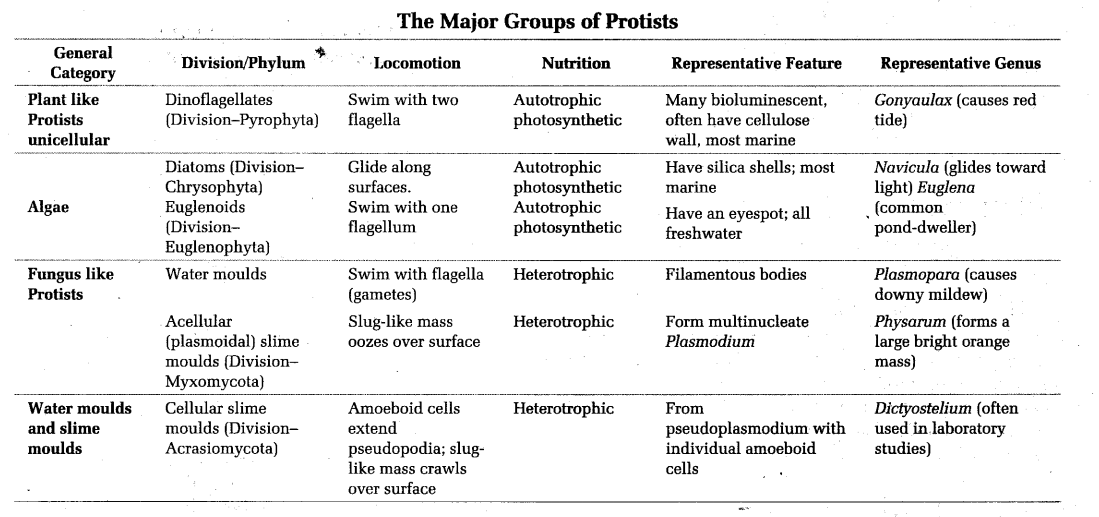
Kingdom-Protista
It includes all the unicellular eukaryotic organisms, e.g. flagellates, diatoms, dinoflagellates, slime moulds, sarcodina, etc. The protists are regarded as ancestors of all multicellular eukaryotic organism.
Phylogenetically, the kingdom-Protista acts as a connecting link between the prokaryotic kingdom-Monera on one hand and the complex multicellular kingdoms of fungi, plants and animals on the other hand.
Special Features of Kingdom-Protista
It includes aquatic organisms, which form planktons. Nutrition is parasitic, ingestive, photosynthetic, saprophytic, etc.
It includes producers, i.e. photosynthetic planktons like algae. It also includes another group of planktons, which is’ non-photosynthetic in nature and is known as zooplankton. It includes Euglena like organism, which has a dual mode of nutrition, i.e. photosynthetic and holozoic (in the absence of light). It also includes organisms, which are intermediate between wall and wall-less organisms, i.e. slime moulds.
Cellular organisation is of two envelopes type, i.e. plasma membrane and internal membrane occur around certain organelles. It possess certain organelles like mitochondria, endoplasmic reticulum, nucleus, food vacuoles, etc. They are motile or non-motile. If motile, they possess flagella or cilia (flagella in (9 + 2) organisation). Reproduction may be sexual or asexual. Cellulose digesting protists occur in termites and wood eating animals.
Kingdom-Fungi
Fungi are a group of eukaryotic, achlorophyllous, non-photosynthetic, heterotrophic and thalloid organisms. The study of fungi is called Mycology. PA Micheli is known as Father of Mycology, whereas EJ Butler is known as Father of Indian Mycology.
Special Features of Kingdom-Fungi
KC Mehta, an Indian scientist is famous for studying rust disease in wheat. When the septa show a pore in the middle of the cross wall, it is known as simple pore (observed in Ascomycetes)..They look like a large jar (the rim is swollen blfrel-shaped guarded by cap-like covers). This is known as dolipore septum (observed in Basidiomycetes). In dolipore septum, the cap is like a round bracket or parenthes. The septal pore cap is called parenthesome. Fungal cell wall consists of fungal cellulose or chitin, a polymer of N-acetyl glucosamine (except Oomycetes, where cellulose occurs as the main component of cell wall).
Reserve food materials found in fungi are glycogen and oil. When a single nucleus present in a cell, the fungal cell is knowm as monokaryon. When two nuclei present in a cell the fungal cell is knowm as dikaryon.
Distribution on the Basis of Mycelium
Most fungi grow as tubular filaments called hyphae. An interwoven mass of hyphae is called mycelium.
When cross walls or septa are formed in the mycelium dividing it into segments, it is known as septate mycelium. Pseudomycelium is a structure observed in yeast, where during budding process, buds are adhered with one another and form a chain-like structure.

Reproduction
At the reproductive stage, the entire cell protoplast (in unicellular lower fungi) is involved in zoospore formation. The coenocytic thallus is known as holocarpic, e.g. Synchytrium.
(i) When the mycelium is differentiated into distinct sterile and fertile portion the thallus is eucarpic, e.g. Phytophthora.
(ii) When two types of strain (+ and -) are required for performing sexual reproduction, the phenomenon is known as heterothallism.
(iii) Blakeslee observed heterothallism in mucorales.
Asexual Reproduction
Zoospores
These are thin walled, motile spores formed in a sporangium. Zoospores may be uniflagellates or biflagellates. The flagella are always heterokont type. Conidia or Conidiophores Spores borne on tips of hyphal structures called conidiophores.
Chlamydospores
These are usually formed during unfavourable conditions and are thick walled, single celled spores, which are highly resistant in adverse conditions,
e.g. Ustilago, Saprolegnia.
Oidia
In some mycelial fungi, the thallus breaks into its component cells. Each cell then round up into a structure called oidium (pi. oidia).
Sexual Reproduction
Sexual reproduction in fungi involves plasmogamy, karyogamy and meiosis. As a result of sexual reproduction, following types of sexual spores are produced:
Ascospores
usually single celled, produce haploid spores in a sac called an ascus,
e.g. Ascomycetes.
Basidiospores
i.e. haploid spores borne on special structures called basidia.
Zygospores
i.e. thick walled diploid spores produced by the fusion of entire gametangia.
The site of meiosis in Oomycetes, Zygomycetes is zygospore/zygote/oospore, whereas ascus in Ascomycetes and basidium in Basidiomycetes.
Economic Importance of Fungi
(i) Yeast (Saccharomyces cerevisiae) is one of the budding yeasts, which ferments sugar to ethanol and carbon dioxide and thus used in
- Making alcoholic beverages like beer and wine.
- Baking (it is often called baker’s yeast). Here, carbon dioxide is used to make bread and cakes rise and have a spongy texture.
- Baker’s yeast is also used in the commercial production of some vitamins and in the production of some human therapeutic proteins by using recombinant DNA technology.
- Yeast is also used as Single Cell Protein (SCP).
(ii) Neurospora is a fungus belongs to
class-Ascomycetes. It is an excellent research material for the researchers in genetics that is why, it is known as Drosophila
- The truffle and the morel are highly priced food delicacies.
- Penicillin is obtained from Penicillium notatum and Penicillium chrysogenum.
(iii) Ashby gosypi, a filamentous yeast is used in the production of vitamin riboflavin.
(iv) Spoilage of food articles such as bread takes place by Rhizopus and Mucor species.
- Mucor arrhizus is used for waste water treatment because it removes heavy metal contamination of water.
- Fermented foods are prepared from rice and soyabean with the help of Rhizopus and Mucor.
- Citric acid is obtained from molasses with the help of Mucor.
- Fumaric acid is obtained from Rhizopus stolonifer.
- Cortisone is obtained by Rhizopus stolonifer.
- Lactic acid is obtained by Rhizopus stolonifer and Rhizopus nodosus.
- Alcohol is obtained by Rhizopus oryzae and Mucor javanicus.
- Other fungi useful for the academic studies are Gibberella fujikuroi (studies on growth regulating substance), Saccharomyces, Aspergillus, etc.
- Bracket fungi or shelf fungi The basidiocarps of these fungi are like brackets or shelves appearing on tree trunk or logs, e.g. Polyporus, Ganoderma, etc.
- Columella is a structure present in sporangium of Mucor.

Mushrooms
These are used as food for their flavour, protein and vitamin contents. Edible part of mushrooms is basidiocarp, which has a fleshy stalk or stipe and umbrella cap or pileus.
Some Edible Mushrooms
- Agaricus campestris (common)
- Agaricus bisporus (cultivated)
- Volvariella volvacea (paddy straw)
- Lentinus edodes (shiitake)
- Armillaria mellea (honey)
- Toadstool A poisonous mushroom is known as toadstool, e.g. Amanita muscaria (fly mushroom).
- Puff-balls These are edible in young state, e.g. Lycoperdon, Clavatia.
- Stinkhorn Produce stinking odour due to the spore mass, which is attractive for flies, e.g. Phallus impudicus (dead man’s finger).Lichens
These are composite organisms, which are formed by a fungus mycobiont and an algal partner called photobiont or phycobiont. This relation is called symbiosis or mutualism. They may grow on rocks, bark, wood, soil, marine or freshwater.
The various types of lichens are:
- Crustose
- Foliose
- Fruticose
- Leprose and
- Filamentous
Algal part prepares food for fungi and fungi provide shelter and absorb mineral nutrients and water for its partner.
Lichens can multiply by fragmentation, isidia and soredia. Sexual reproduction and asexual spores are formed only by the mycobiont.They are sensitive to sulphur dioxide and are hence, the indicator of pollution. They do not grow in polluted areas. Lichens are highly sensitive to air pollution.
Mycorrhiza
It is an association between a fungus and the root of a higher plant, e.g. pine birch. Mycorrhizal roots occur in superficial layers of soil. Mycorrhiza is of two types, i.e. ectomycorrhiza and endomycorrhiza. Mycorrhiza is an example of symbiosis or mutualism. The fungus obtains shelter and food from foot. It helps the root in absorption of water, dissolution and absorption of inorganic nutrients locked in organic matter (especially nitrogen and phosphorus) and protection from other fungi.